
Cell Anatomy Vector Illustration Labeled Educational Structure Diagram Stock Illustration
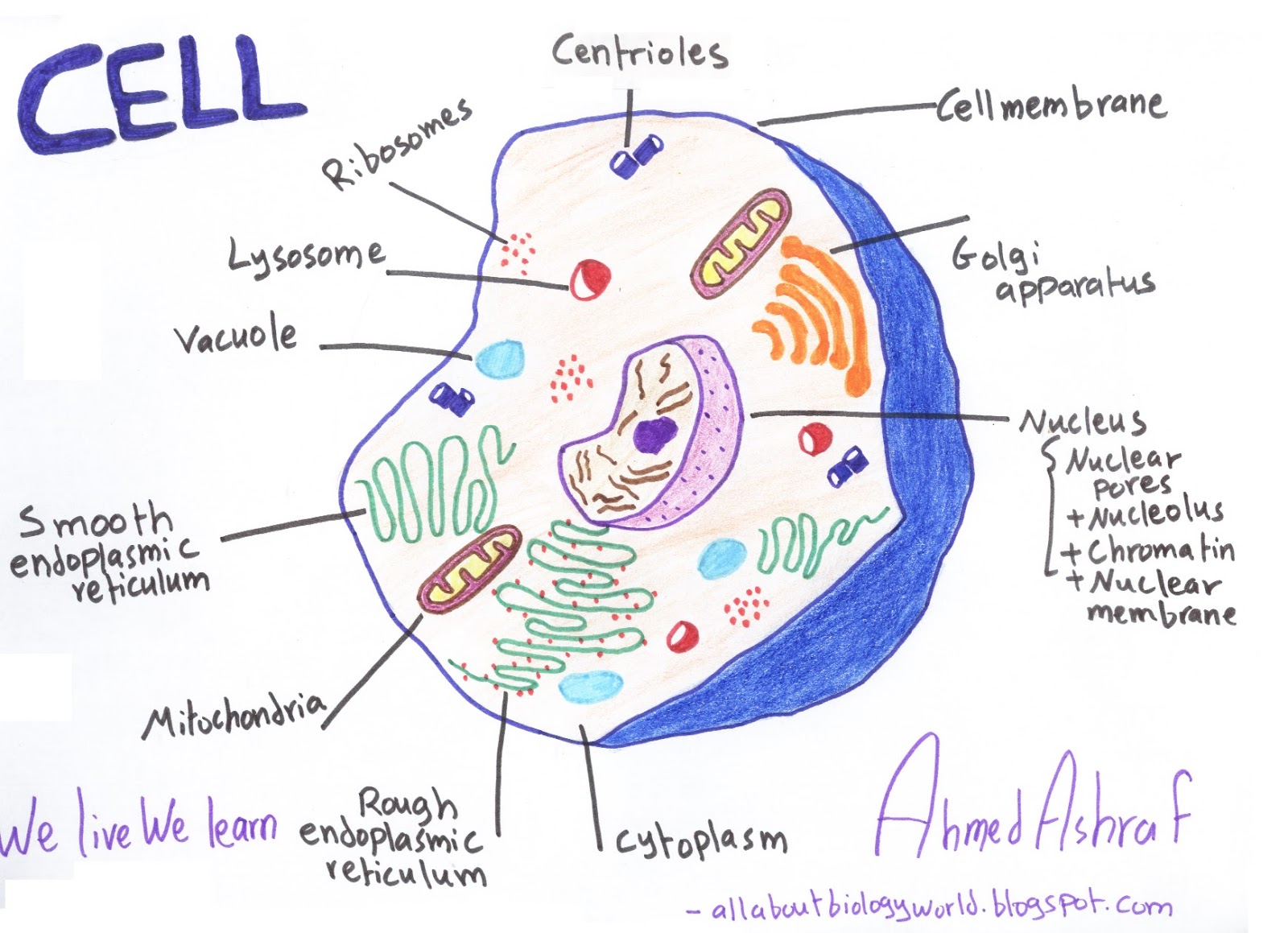
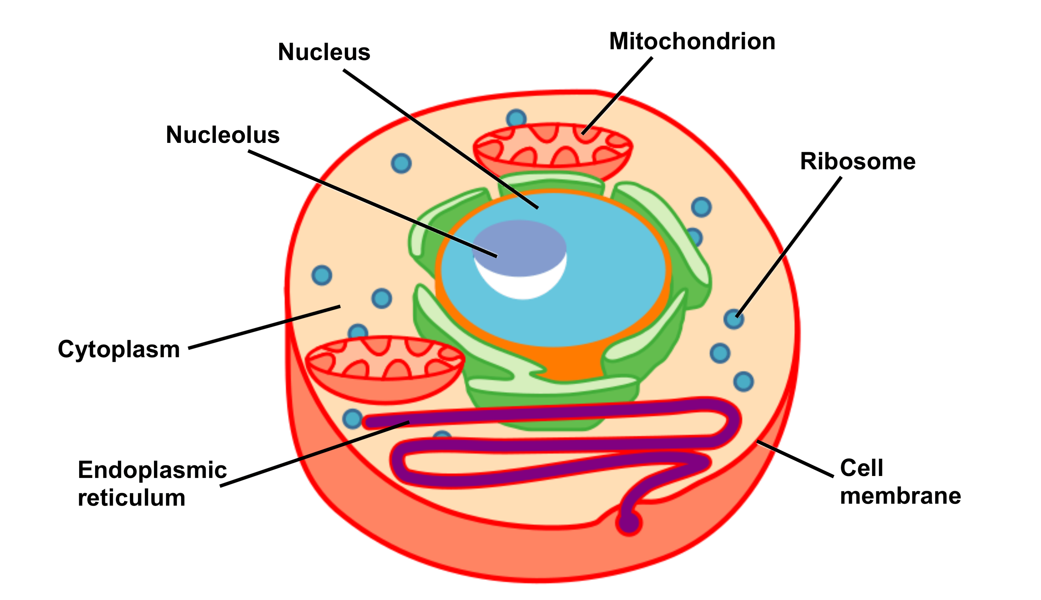
1. Draw a simple circle or oval for the cell membrane. The cell membrane of an animal cell is not a perfect circle. You can make the circle misshapen or oblong. The important part is that it does not have any sharp edges. [1] Also know that the membrane is not a rigid cell wall like in plant cells.
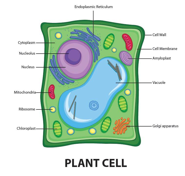
Draw a welllabelled diagram of a plant cell.
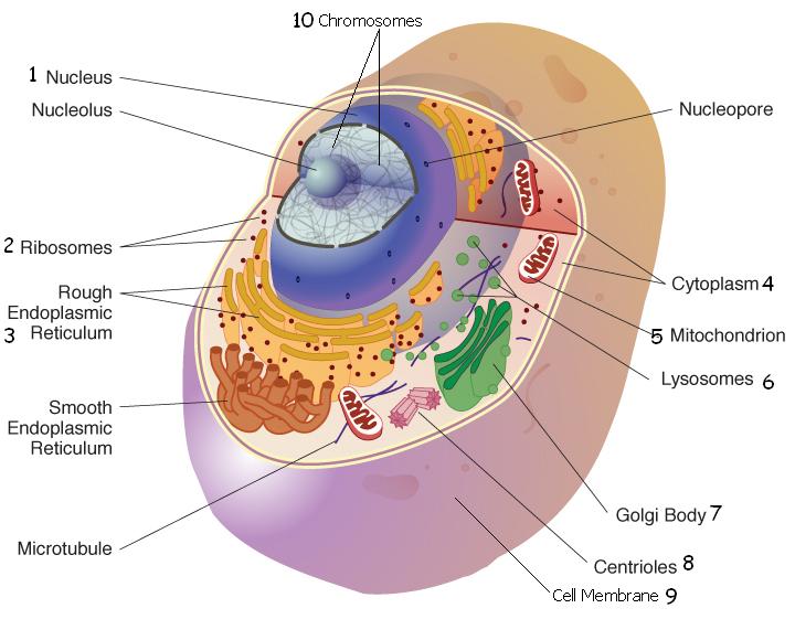
Diagram Of Animal Cell. Animal cells are eukaryotic cells that contain a membrane-bound nucleus. They are different from plant cells in that they do contain cell walls and chloroplast. The animal cell diagram is widely asked in Class 10 and 12 examinations and is beneficial to understand the structure and functions of an animal.

Label The Parts Of A Cell
Labeled Animal Cell Diagram. Blank Animal Cell Diagram Worksheet. The third and fourth diagrams are animal cell diagram worksheets. Quiz yourself by filling in the blanks. Unlabeled Animal Cell Diagram. Finally, an unlabeled version of the diagram is included at the bottom of the page, in color and black and white. This may be useful as a.

Biology 101 Cells Owlcation
cell, in biology, the basic membrane-bound unit that contains the fundamental molecules of life and of which all living things are composed.A single cell is often a complete organism in itself, such as a bacterium or yeast.Other cells acquire specialized functions as they mature. These cells cooperate with other specialized cells and become the building blocks of large multicellular organisms.

Plant Cell Parts and Structure
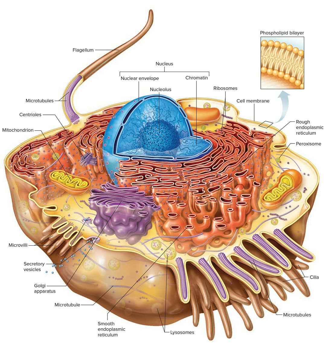
7 years ago. The Endoplasmic Reticulum in a eukaryotic cell is the transport network of the cell and it extends from and connects the nuclear membrane to the plasma membrane of a cell. But then whenever we draw a diagram of a typical plant or animal cell, we never extend it to the plasma membrane- we always leave it somewhere in the cytoplasm.

South Pontotoc Biology Plant and Animal Cell Diagrams
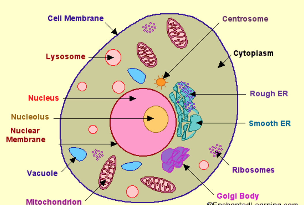
The nucleus is a large organelle that contains the cell's genetic information. Most cells have only one nucleus, but some have more than one, and others—like mature red blood cells—don't have one at all. Within the nucleus is a spherical body known as the nucleolus, which contains clusters of protein, DNA, and RNA.

Animal Cell diagram with labels by Russell Kightley Media
In animal cells, there are many small ones (rather than one large one in plant cells). They are circular in shape. The command center of the cell. It controls cell's activity. The storage facilities for the cell. They store food or waste. Plant cells only have one and it takes up about 90% of plant cell.
Human Cell
Animal Cell: Structure, Parts, Functions, Labeled Diagram. June 6, 2023 by Faith Mokobi. Edited By: Sagar Aryal. An animal cell is a eukaryotic cell that lacks a cell wall, and it is enclosed by the plasma membrane. The cell organelles are enclosed by the plasma membrane including the cell nucleus. Unlike the animal cell lacking the cell wall.
[DIAGRAM] Parts Of A Cell Diagram
Animal Cell Anatomy. The cell is the basic unit of life. All organisms are made up of cells (or in some cases, a single cell). Most cells are very small; in fact, most are invisible without using a microscope. Cells are covered by a cell membrane and come in many different shapes.

cell structure Animal cell drawing, Cell diagram, Human cell diagram
We have included three variations of this worksheet, including. Animal Cell - Labeled for use as an anchor chart or note-taking worksheet. Animal Cell Labeling Diagram - Fill in the blanks to label animal cell structure. Animal Cell Coloring Sheet - Label the animal cell diagram by following the color code to identify animal cell structures.
Biology Club Our cells 1 ( structure, function, division, disorder & cycle )
An animal cell diagram with labels provides a visual representation of the different organelles, such as the nucleus, mitochondria, endoplasmic reticulum, Golgi apparatus, and lysosomes. Labels help to identify these organelles and understand their specific roles in cellular processes like protein synthesis, energy production, and waste disposal.

Plant Cell Diagram, Definition, Structure, Function & Parts
A diagram of a cell with labels is a visual representation of the different parts and structures within a cell. It provides an overview of the intricate organization and complexity of cells, which are the basic building blocks of all living organisms. By understanding the diagram and the labels associated with each component, we can gain.
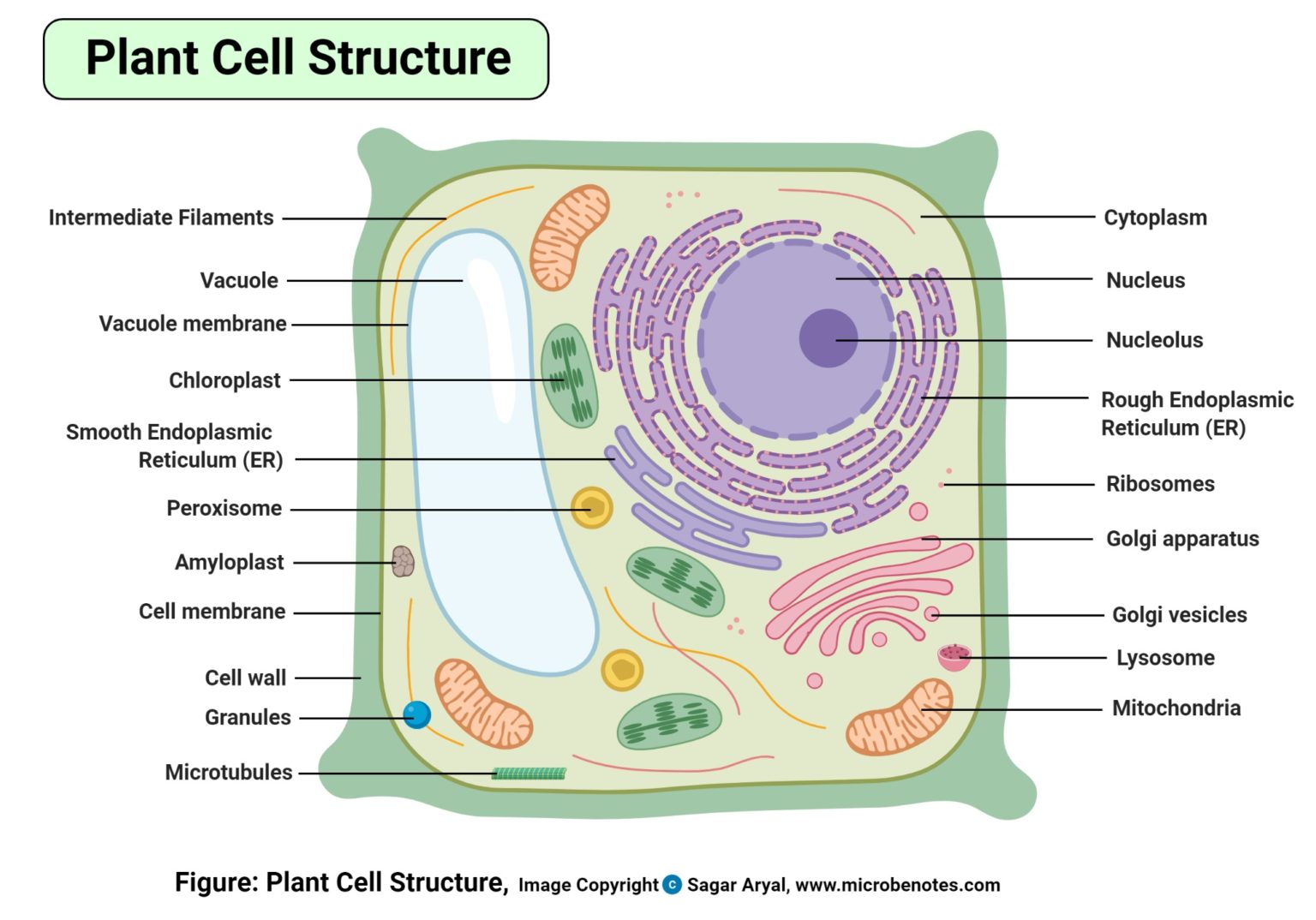
Plant Cell Structure, Parts, Functions, Labeled Diagram
Cell diagram labeled. For this exercise we'll start with an image of a cell diagram ready labeled. Study this and make sure that you're clear about which structure is found where. Cell diagram unlabeled. It's time to label the cell yourself! As you fill in the cell structure worksheet, remember the functions of each part of the cell that.
Cell Structure and Function Part 1 The Organelles Medical Exam Prep
A medium-sized circular cell part that has squiggly lines inside is labeled nucleus. The outermost part of the cell, which is shown as an outline of the cell, is labeled cell membrane. On the right is a four-sided figure with rounded corners that represents a plant cell. The cell contains many cell parts with different shapes.
Cell Biology, Cell Structure
Cell Diagrams with Labelling Activity. I've created two interactive diagrams for an upcoming open textbook for high-school level biology. The cell structure illustrations for these diagrams were generated in BioRender. Both diagrams feature a drag-and-drop labelling activity created with H5P here on Learnful.

Plant Cell Diagram Labeled Class 9 Labeled Functions and Diagram
A cell is the smallest living thing in the human organism, and all living structures in the human body are made of cells. There are hundreds of different types of cells in the human body, which vary in shape (e.g. round, flat, long and thin, short and thick) and size (e.g. small granule cells of the cerebellum in the brain (4 micrometers), up to the huge oocytes (eggs) produced in the female.